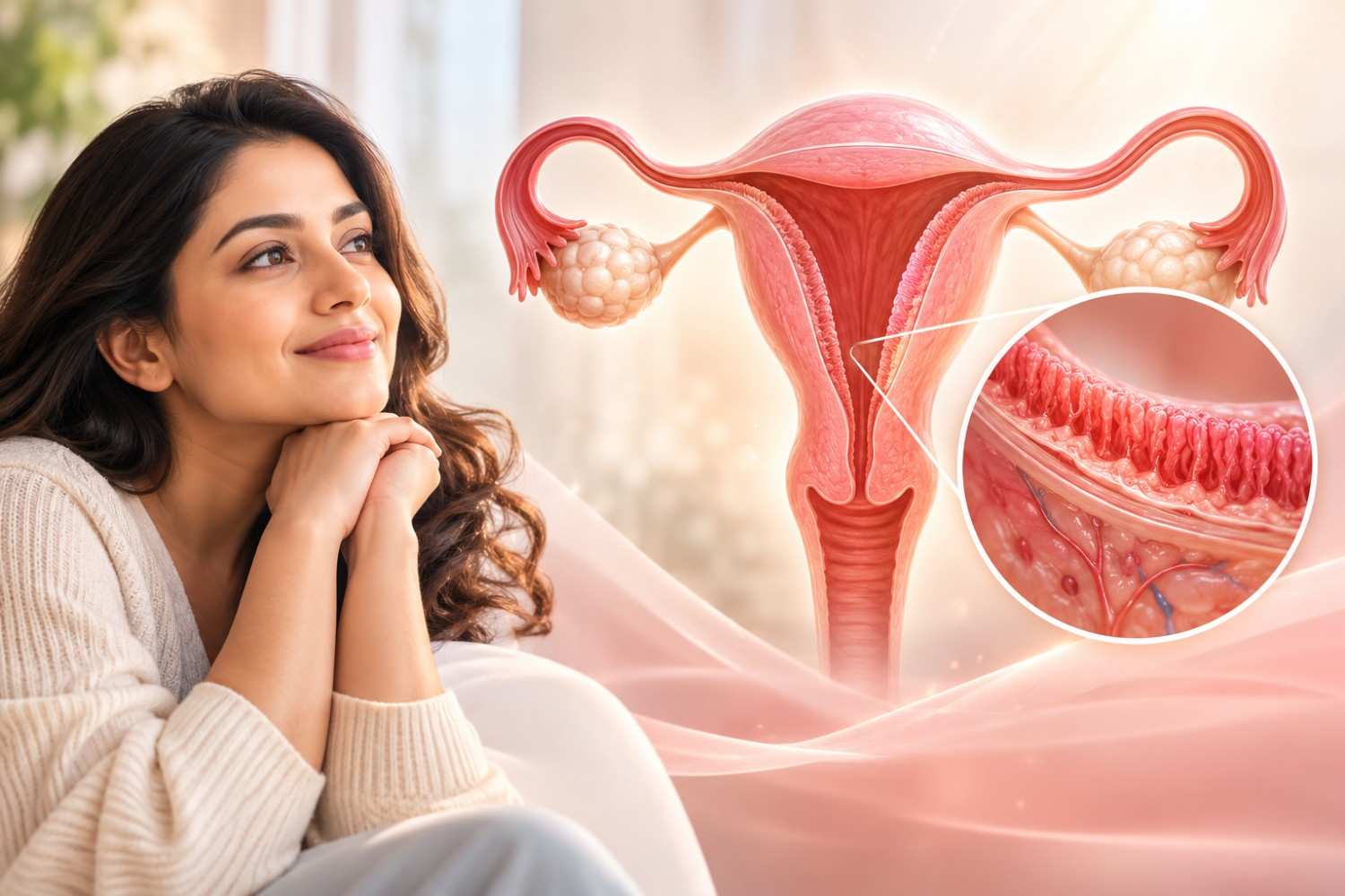
A thin uterine lining is one of the more frustrating findings in a fertility work-up. You may be having regular cycles and good follicle growth, yet be told that the lining of the womb is not developing as well as hoped. The good news is that a thin endometrium has identifiable causes in many women, and there are practical ways to evaluate and manage it.
This page explains what a thin endometrium means, why it happens, how it is assessed and the treatment options available. It is general educational information. The right plan depends on your age, history, reports and fertility goals, and can only be decided after a personal consultation.
The endometrium is the inner lining of the uterus. Each month, under the influence of hormones, it thickens to prepare for an embryo to implant. After implantation, it nourishes the early pregnancy.
Lining thickness is measured by ultrasound. Many fertility specialists consider a lining below about 7 mm around the time of ovulation or embryo transfer to be thin, although the exact cut-off varies between clinics and studies. Pattern and blood flow matter as well as thickness. A thin lining does not mean pregnancy is impossible, but on average it is associated with a lower chance of implantation, so it deserves attention.
Many women have no symptoms and the problem is found on a routine fertility ultrasound. You should mention the following to your doctor:
A thin endometrium has identifiable causes in many women, and understanding the underlying reason helps guide evaluation and management.
Oestrogen drives growth of the lining. Low levels, poor ovarian response, premature ovarian insufficiency or thyroid and other endocrine problems can limit it.
The lining depends on a good blood supply. Reduced uterine blood flow may be seen in some women and can be assessed with Doppler ultrasound.
Repeated or aggressive uterine procedures such as dilatation and curettage (D&C), surgery for retained products after miscarriage or delivery, or infection can cause scar tissue inside the uterus. Scarring can leave too little healthy lining to respond to hormones, and is often associated with very light or absent periods.
Long-standing inflammation or infection of the lining, including genital tuberculosis in some women, can damage the endometrium and affect its growth.
Certain ovulation-inducing medicines, notably clomiphene citrate, can have an anti-oestrogen effect on the lining in some women, even when ovulation occurs well.
Fibroids or polyps that distort the cavity, previous pelvic radiation, advancing age and, in some cases, no clear cause can also be associated with a thin lining.
The aim is to measure the lining accurately and, more importantly, to find out why it is thin.
Your doctor will ask about menstrual flow, previous pregnancies, miscarriage or abortion procedures, uterine surgery, infections, medicines and previous fertility treatment.
Lining thickness and pattern are checked at specific times in the cycle, usually in the late follicular phase near ovulation, or before an embryo transfer. Serial scans may be needed because thickness changes through the cycle. Doppler studies can assess blood flow, and a 3D scan can show the cavity in more detail.
Hysteroscopy allows the inside of the uterus to be viewed directly with a thin camera. It is the most reliable way to identify adhesions, polyps and other cavity abnormalities, and treatment can often be done in the same procedure.
Depending on your history, blood tests such as thyroid function, prolactin, FSH, oestradiol and AMH may be advised. If infection or chronic endometritis is suspected, an endometrial sample may be examined or tested, including for tuberculosis where clinically appropriate.
Treatment is directed at the cause and tailored to the individual. There is no single treatment that works for every woman, and some commonly discussed options have limited or mixed evidence. A responsible plan explains this clearly.
If hysteroscopy shows adhesions, a polyp or a septum, these can be treated with hysteroscopic surgery. After adhesion surgery, hormonal therapy and follow-up are often used to support healing. Infection or chronic endometritis is treated with appropriate medicines after testing, and thyroid or other endocrine problems are corrected.
Oestrogen given as tablets, patches or vaginal preparations may be used to encourage lining growth, with the dose and duration individualised and monitored by ultrasound. This is commonly used in cycles planned for frozen embryo transfer.
If a medicine used for ovulation induction appears to be thinning the lining, your doctor may change the drug, dose or protocol. In IVF, embryos can be frozen and transferred in a later, hormonally prepared cycle when the lining is better, rather than in the same stimulation cycle.
Some clinics use low-dose aspirin, vaginal sildenafil, pentoxifylline with vitamin E, or other measures to try to improve blood flow, and newer approaches such as platelet-rich plasma (PRP) or G-CSF have been explored. Evidence for these is limited and results vary, so they should be considered only after a frank discussion of potential benefits, uncertainty and side effects, and not as guaranteed solutions.
Staying active, avoiding tobacco, maintaining a healthy weight, managing stress and keeping medical conditions under control support general reproductive health. These measures are helpful but are not a substitute for treating a specific cause.
If the lining does not respond despite appropriate treatment, your doctor will discuss realistic options with you, which may include continuing monitored attempts, further evaluation, or, in selected situations, other family-building routes. Decisions are made together, at your pace.
At your first visit, your history and previous reports will be reviewed, and a pelvic ultrasound may be performed. You will be told which further tests are needed and why, what the findings mean, and what treatment options are available, including their benefits and limits.
Please bring previous scans, hysteroscopy or surgical notes, hormone reports, and details of earlier treatment cycles. If you are married, attending with your partner helps the doctor plan fertility care for both of you.
Dr. Sunita Arora is an obstetrician, gynaecologist and fertility specialist with more than 23 years of experience. She holds an MBBS from Lady Hardinge Medical College, New Delhi, and an MD in Obstetrics and Gynaecology from UCMS and GTB Hospital, and she is a Fellow of the Indian College of Obstetricians and Gynaecologists (FICOG) and a Fellow of the International College of Surgeons (FICS). She completed a fellowship in Reproductive Medicine at D.Y. Patil University, Mumbai, and received IVF training at RMA, Mount Sinai, New York.
Her areas of interest include infertility, IUI, IVF and endoscopic surgery, which are directly relevant to the evaluation and management of uterine factors.
If a thin uterine lining has been mentioned in your scan reports, or you have had difficulty conceiving or failed treatment cycles, a careful evaluation can bring clarity.
She consults at Safdarjung Enclave and is also associated with Fortis La Femme, Greater Kailash II, serving women across South Delhi, including Greater Kailash, Green Park, Hauz Khas and nearby areas.
Dr Sunita Arora Fertility and Gynae Clinic
Fortis La Femme, South Delhi
Book an appointment with Dr. Sunita Arora to review your reports and plan the next steps that are right for you.
Find answers to common questions about thin endometrium causes, diagnosis, and treatment options.
Medical disclaimer: This page is for general educational purposes and does not replace personalised medical advice, diagnosis or treatment. Please consult a qualified doctor about your own health. If you have severe abdominal pain, heavy bleeding, fainting or fever, seek urgent medical care.